I don't know if anyone did Medical Physics but I'm having a hard time understanding the difference between T1 & T2 weighted for MRI.
Can anyone distinguish this for me?
Hey! It was my option (Jake's too), happy to lend a hand! This was probably the most difficult concept in the course for me, so I hope I can explain it in a way that is beneficial

Okay, so we know that when we expose nuclei to radio waves of an appropriate frequency, they resonate and
precess, and this causes change to the
net magnetisation vector. Relaxation is related to this vector.
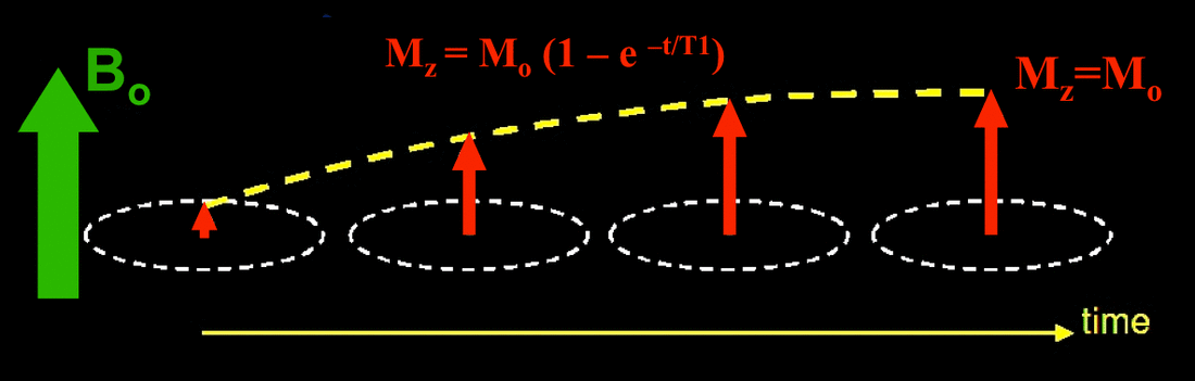
T1 Relaxation: As nuclei are exposed to radio waves, they flip into anti-parallel alignments. This shrinks the component of the net magnetisation vector in the direction of the file. T1 Relaxation concerns this vector returning to its initial value (specifically, the T1 relaxation time is when the vector returns to 63% of the initial value), as nuclei dissipate their energy into the surrounding lattice. This diagram does a pretty good job showing what this looks like (ignore the equation, it's beyond this course):
 T2 Relaxation
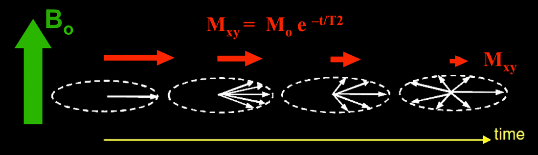
: For reasons slightly beyond the scope of the courses, exposing precessing nuclei to radio frequencies causes the net magnetisation vector to shift into the transverse plane, perpendicular to the applied field. Remember that previously, there was no component of the vector in this direction, it was only parallel to the field. The result is that the precession of the nuclei as a whole loses coherency and phase. As the nuclei relax, those precessing about this transverse axis transfer their energy to those precessing parallel to the field, and the component of the vector perpendicular to the field shrinks. T2 Relaxation Time is when the vector shrinks back to 37% of its initial. Again, see diagram:
That is the complicated version: If you want simple, go for this instead. Applying a magnetic field rotates the net magnetisation vector 90 degrees, so it ends up perpendicular to the applied field. T1 Relaxation time measure the parallel component increasing to normal, T2 relaxation time concerns the perpendicular component shrinking away. This is kind of why the two numbers (63% and 37%) are related (adding to 100%), because they concern the same vector

I hope this makes sense! Understanding this demands a rock solid knowledge of everything else in this section; and then a lot of dedication and research to really wrap your head around it!
Oh! So for actually weighting to detect these relaxation times, a few things to note about MRI imaging radio waves first.
Repetition Time (TR): Elapsed time between pulses of radio waves
Echo Delay Time (TE): Time delay between the sending of radio waves and measurement of emitted signals
For T1 weighted images, we want to emphasise areas with short T1 relaxation time (meaning, they dissipate energy very quickly). This is achieved by minimising the repetition time, meaning that only nuclei with short T1 will have the time to dissipate their energy before the next pulse.
For T2 weighted images, we want to emphasise areas with a long T2 relaxation time (meaning, it takes a long (relative) time for them to return to their initial coherency, and thus, will emit MR signals for longer). We do this by maximising echo delay time, so that by the time we take the measurement, only nuclei with long T2 will still be emitting MR signals
